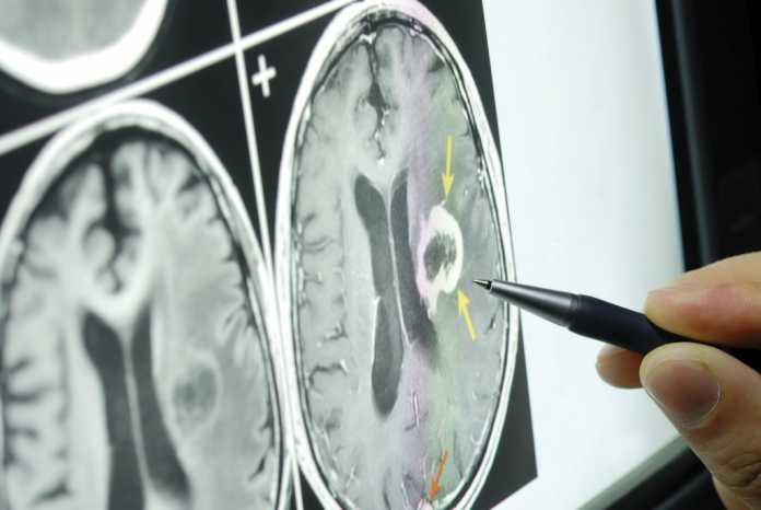
Brain tumor diagnosis is very nice topic discussed. Dr. Len Cerullo, neurosurgeon, discusses the diagnostic tools used by neurosurgeons to reveal the size, location and nature of a brain tumor. The various types of imaging studies are discussed in detail, including MRI, CT scan, MR spectroscopy, functional MR. Dr. Cerullo also discusses the use of physiological testing, including visual field testing, brain stem evoked potential testing and somatosensory evoked potential testing.
Brain tumor diagnosis
We’re going to talk about how we look at the brain in people who are suspected of having a brain tumor and of course the level of suspicion move determined to some degree the timing and the type of test in the old days we were constrained. We can do an x-ray of the skull looking for a shift of the pineal gland, which is a small calcification right in the center of the head and if the pineal we shifted to one side. Then we knew that the tumor was on the other side and vice versa.
And we have angiography which was a way to look at the blood vessels of the brain, and by determining where those blood vessels were deformed or draped or abnormal, we could get a kind of an idea about where the tumor is and all that changed. In the late 60s early 70s when CT scanning became available and CTS getting at that time was about as sophisticated as a handheld computer would be today, but it was so much better than anything we had had to work with for time immemorial before shortly after CT scanning, which looks at the density of the brain. Every investigation should be coincides with symptoms of brain cancer.
In other words, the the structures which are more dense look white on a CT scan stretches, which are less dense like fatter fluid black on a CT scan, but shortly after that we have the opportunity to use MRI scanner magnetic resonance imaging MRI, and this is a very, very good way to look at the soft tissues, and it looks at the water content of the soft tissues and by enhancing the study injecting a Diet Coke gadolinium we can get a very, very good impression. Not only abnormal tissue but of abnormal tissue its location its relationship to healthy tissue. The Associated effects. It is fluid on the brain or hydrocephalus fluid within the tissue or demon. By the way, here is 10 symptoms of brain tumors you should never ignore.
So the MRI scan today would be considered the gold standard for brain tumors. It’s not the only study that can be for or even should be performed, but it is the best starting place. And that’s why today we are seeing so many patients with what we call incidental tumors or tumors that weren’t even suspected clinic believe, but they showed up on the MRI scan and then, of course, the question is what to do, and that’s a very challenging area of Neurosurgery and also medical ethics at any rate.
MRI scan is performed in a kind of a donut in a tube, and the open scans are a little bit freer the clothes scans are relatively claustrophobic and about 10% of people will need to have some sedation, just to tolerate the skin because they’re kind of in this closed space which is likened to being in a coffin that that bed but it is locked. People sometimes need sedation, and at any rate, the molecules of the brain, the water molecules in the brain are lined up by magnetic fields and in zapped with a radio frequency currents of the spin down to their resting state in spinning down they give off energy and the energy is picked up by the sensors throughout the entire periphery and then complex calculations are performed and at the end of the day, we have a picture of the brain.
We have a view of the mind in this direction which is called the axial direction and this direction which is called this coronal direction and right in this direction which is called the satchel direction and then these can be further manipulated to create 3D images or to rotate the images in certain ways. And it’s a very sophisticated to which gives us a very, very good picture of what’s going on.
Then depending on the type of lesion or two, we may look to other types of scanning to confirm or deny the presence of blood. The presence of calcifications the metabolic activity of the brain in the tumor and surrounding the tumor the location of the tumor visa V functional areas that are called Functional Mr.
And that’s used when the lesion is in an area which may control speech so that the MRI scan is done with certain other computer protocols with certain different types of chemicals and we can determine by asking the patient to speak or to read or to write during the scan exactly where those functional areas of the brain are these are the bad areas the tumor areas magnetic spectroscopy is another type of Mrs skin which shows us the chemical balance within the tumor itself and the chemical balance in the tumor is usually different especially in malignant tumors. It’s different from the normal surrounding brain. So if we have kind of an abnormal area we can on the basis him MR spectroscopy, get some appreciation of whether indeed this is tomorrow or this is scar tissue or this is a low-grade tumor or something else.
PET scan positron emission tomography of the brain has also been used primarily in recent years, and especially in people who have tumors elsewhere in the body. So if we’re looking through a big area like a whole body. We can do a PET scan and find something in the brain, or if there’s some question whether the abnormality we saw on the MRI scan of the brain is really a tumor or maybe just scar tissue. We can sometimes do a PET scan, and that like MR spectroscopy will help to differentiate between the two. At the end of the day for most tumors.
The best diagnostic treatment is to take a piece of the tumor and look at it under the microscope and analyze its DNA and that’s the best way to identify precisely what it is and hopefully by identity just what it can plot a course of action, whether that be chemotherapy or radiation therapy or genetic manipulation therapy or all of the above.
And that’s where we are with imaging of the brain, the imaging of the brain is a graphic descriptive term.
We also have physiologic tests that can be done to look at different areas of the brain. So if the tumor is located in around any of the visual pathways from the eyeball all the way back to the occipital lobe. We will do a visual field examination which plots out the visual fields of each eye and enables us to identify where in that visual pathway deletion is. Similarly, if we’ve got a tumor or a lesion along the brain stem or the nerve to the hearing have the nerve to facial movement of the facial nerve sensation, and we do brain stem a vote testing and if we have a tumor lower in the brain stem which may affect the feeling coming up from the body.
We do submit a sensory evoked potential testing. These tests are also useful in that they can be used during surgery to enhance the safety of the operation and the efficacy of the operation. So we have physiological ways to look at the Ring anatomical or radiographic ways to look at the brain, and between the two we get quite a good picture of what’s going on and what we can do about it all the way from the few cell level to the zillion of cell level that release but billion.


